Diagnostic Imaging Tools Used to Evaluate Your Joint Pain
What is a CT scan? What does an MRI do? In this article, Orthopedic Surgeon Dr. Sperling discusses common types of diagnostic imaging used in diagnosing joint pain.
What is a CT scan? What does an MRI do? In this article, Orthopedic Surgeon Dr. Sperling discusses common types of diagnostic imaging used in diagnosing joint pain.
Over the past 40 years, there have been dramatic advances in the field of radiology that have improved a clinician’s ability to evaluate the status of soft tissue and bone structures in a patient’s joint. For many years, plain radiographs, or X-rays, were the only imaging technology available. This provided limited information for understanding potential causes of joint pain outside of a physical examination and the history of discomfort.
The development of new imaging techniques such as magnetic resonance imaging (MRI), computerized tomography (CT scans), and ultrasounds have significantly improved the ability of your physician to not only diagnose what could be causing your joint pain, but also help guide specific treatments.

Plain radiography (X-ray)
Plain radiographs, better known as X-rays, provide important information for your surgeon to help evaluate your joint and the potential sources of pain that you’re experiencing. Radiographs show the presence of joint space narrowing that is seen with joint pain due to arthritis. For example, the bone in your shoulder joint is lined with cartilage, a smooth lining that allows for smooth motion. The cartilage in your shoulder is similar to the rubber on a tire of a car. Your surgeon can see on the X-ray whether the joint has been worn down, similar to the rubber on a tire being worn down over time. Your surgeon can also get a sense of how much bone has been worn away due to the arthritic process.
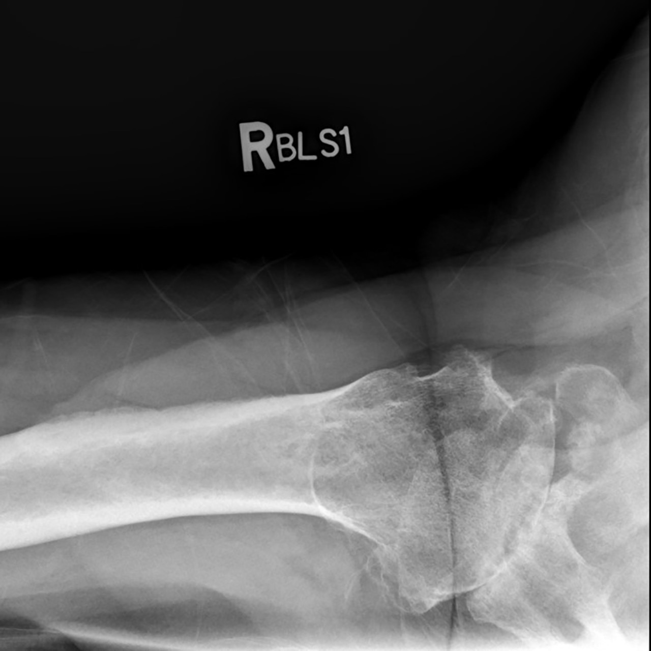
Figure 1: Plain radiograph example of bone-on-bone arthritis with wear of the socket
Osteophytes, or perhaps better known as bone spurs, can also be seen on plain X-rays. Osteophytes can be another sign of arthritis. Rather than the ends of your joint being round, they begin to flatten which can also be seen on the plain radiographs. Sometimes, the surgeon can obtain an overall picture of bone quality. Lastly, radiographs provide important information on whether the alignment of the joint looks abnormal. In some types of arthritis, there may be subluxation, or shifting of the joint surface, which can be another sign of joint wear.
Ultrasound
The technology behind an ultrasound has undergone a significant revolution over the past 10 years. Ultrasound machines have become smaller and have improved your doctors ability to visualize your joint. The real power of an ultrasound is that it can assess the joint in motion… it’s dynamic. An ultrasound can be a convenient and in-office technique used to evaluate soft tissue structures inside your joint as well.
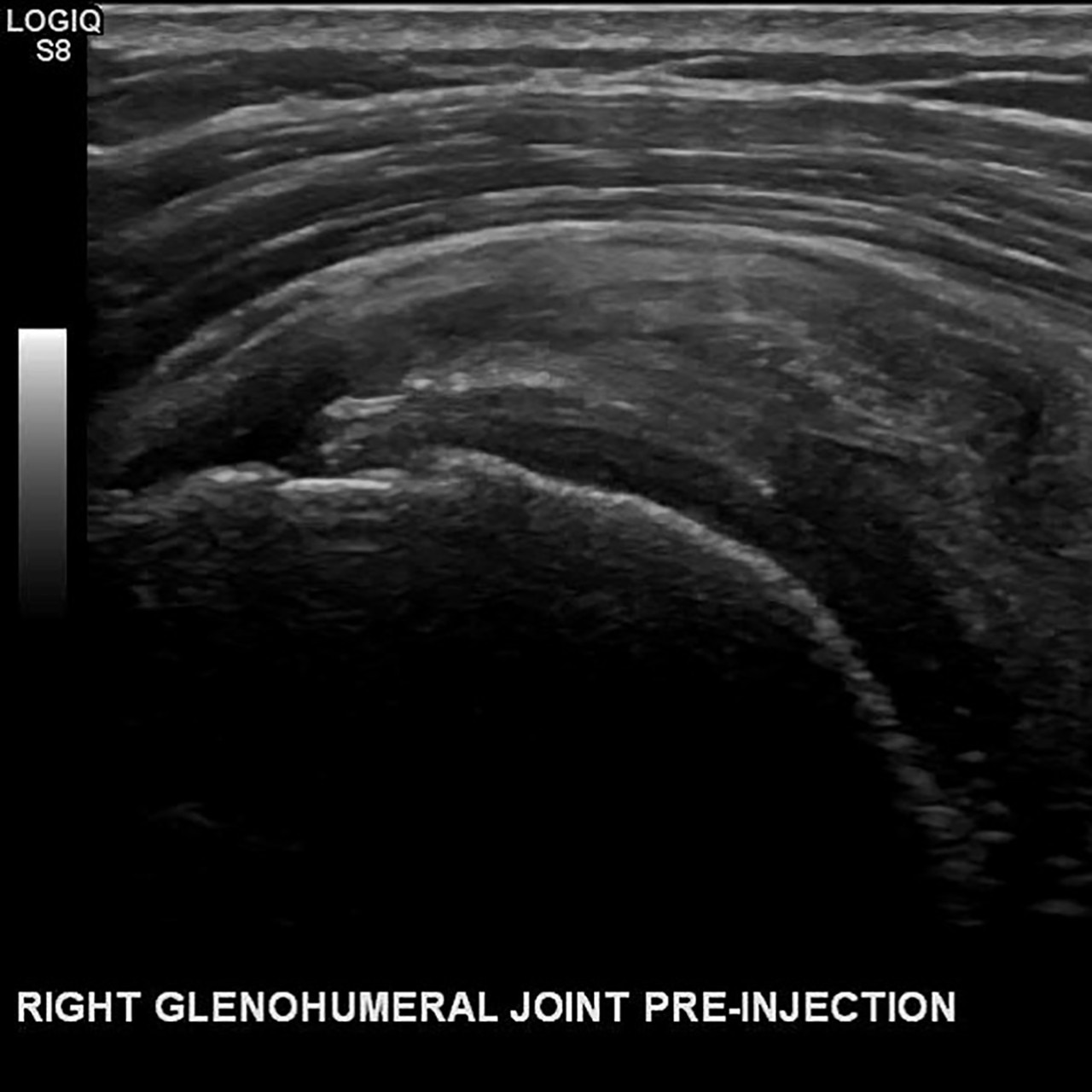
Figure 2: Ultrasound image of the rotator cuff and tendon
Ultrasounds can be used to help the placement of needles within the joint for both diagnostic and therapeutic purposes. Sometimes, your doctor may use the ultrasound machine to confirm the exact placement of a needle used for an injection. The doctor can also use the ultrasound machine to confirm placement of the needle in a specific location for aspiration-or withdrawing fluid for testing.
Magnetic resonance imaging (MRI)
Magnetic resonance imaging (MRI) is one of the most powerful diagnostic tools available. The developers of the MRI won the Nobel Prize in 2003 for their tremendous advancement and impact on patients around the world. Unlike X-rays or CT scans, MRI machines do not use radiation.
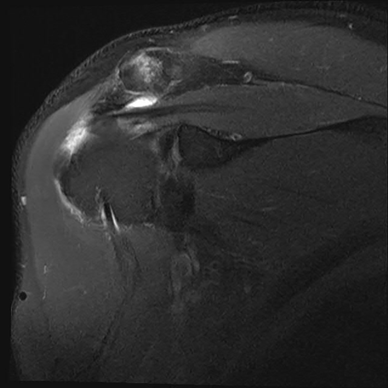
Figure 3: MRI image of a torn rotator cuff
The great strength of an MRI is the ability to visualize soft tissue within your joint. An MRI may be performed with or without contrast based on the preference of your doctor and the specific objectives of the MRI. The quality of MRI continues to improve significantly and now your surgeon can gain greater clarity on what may be occurring inside the joint causing your pain. An MRI has the ability to detect not only full thickness tears of structures (such as your rotator cuff in the shoulder), but also subtle partial tears.
Computerized tomography scan (CT scan)
A computerized tomography, otherwise known as a CT scan, uses rotating X-rays and computers to create images of your joint. CT scans provide great detail of your joint, particularly related to the bone. Thin cut CT scans are becoming more commonly used among some surgeons in planning joint replacement surgery. These scans can provide great detail of the exact contour of your joint. Compared to an MRI, a CT scan is usually faster and requires less time for you to be in the scanner. A CT scan may be performed with or without contrast based on the specific diagnostic goals of your physician.
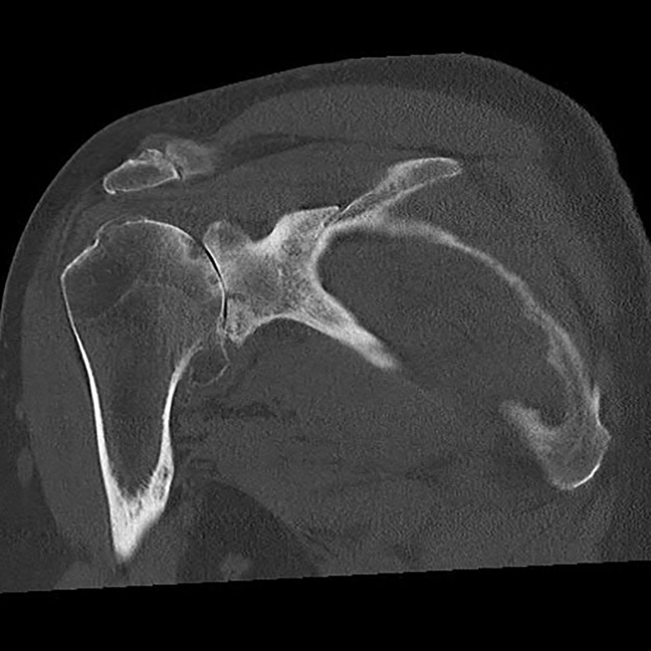
Figure 4: CT scan demonstrating bone-on-bone shoulder arthritis with wear of the socket